Jezek 2014 Abstract MiP2014
| Mitochondrial cristae morphology changes: from Hackenbrock to hypoxia. |
Link:
Mitochondr Physiol Network 19.13 - MiP2014
Jezek P, Engstova H, Alan L, Spacek T, Stradalova V, Malinsky J, Bewersdorf J, Dlaskova A, Plecita-Hlavata L (2014)
Event: MiP2014
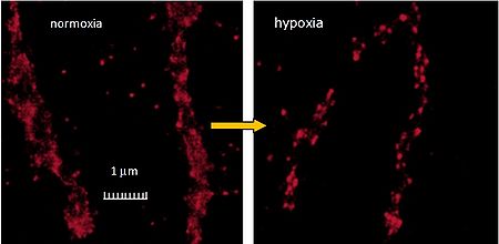
Hackenbrock’s classic observation [1] distinguished orthodox cristae at a resting LEAK state and condensed cristae conformation for isolated mitochondria at the active phosphorylating OXPHOS state, which are, however, not encountered in situ [2]. Since it resides in MINOS complexes, joining the mitochondrial inner membrane, mtIM, and outer membrane (mtOM), the mtIM protein mitofilin contributes to cristae shapes. The aim of our study was to find out how altered metabolism at hypoxia is reflected by changes in cristae morphology. 3D dSTORM microscopy (3D immunocytochemistry) showed distinct mitofilin foci projected on the mtOM in HepG2 cells cultivated for 72 h at 5% oxygen (termed "hypoxic cells"), while the mtOM-projected surface density of mitofilin molecules vs. normoxic cells decreased by ~40% (Fig. 1), accompanied by ~20% loss of mitofilin and its transcript. Cryo–electron microscopy documented intracristal space (ICS) expansion by cristae width increase, predominantly in glycolytic cells, not occurring in reduced or mtOM–detached cristae of OPA1– and mitofilin–silenced HepG2 cells, respectively. The hypoxic ICS expansion resembles Hackenbrock’s classic observation of condensed cristae [1]. Moreover, we confirmed paradoxical observations of orthodox cristae in cells undoubtedly phosphorylating, which had more shrunken ICS and expanded matrix space at atmospheric oxygen. In turn, upon hypoxia, the IMS expansion reflected the established Hackenbrock condensed cristae conformation [1]. Furthermore, ATP-synthase dimers vs. monomers ratio and OXPHOS/LEAK respiratory control ratios were higher under normoxia. Since these ATP-synthase dimers predominantly locate at sharp cristae edges, whereas at hypoxic ICS expansion these edges are disrupted at more round cristae, we hypothesize that for glycolytic cells the ICS expansion represents adaptation, decreasing the number of ATP-synthase dimers, serving for ATP synthesis downregulation during cell survival under hypoxia.
• O2k-Network Lab: CZ Prague Jezek P
Labels: MiParea: Respiration, mt-Structure;fission;fusion, mt-Membrane
Stress:Ischemia-reperfusion
Tissue;cell: Other cell lines Preparation: Isolated mitochondria
Regulation: ATP production Coupling state: LEAK, OXPHOS
HRR: Oxygraph-2k Event: A2, Oral MiP2014
Affiliation
1-Inst Physiol; 2-Inst Experim Medicine, Acad Sc, Prague, Czech Republic; 3-Dep Cell Biol, Yale Univ, New Haven, CT, USA. - [email protected]
Figure 1
References and acknowledgements
Supported by GACR grants P302/10/0346 and 13-02033S.
- Hackenbrock CR (1966) Ultrastructural bases for metabolically linked mechanical activity in mitochondria. I. Reversible ultrastructural changes with change in metabolic steady state in isolated liver mitochondria. J Cell Biol 30: 269–97.
- Sun MG, Williams J, Munoz-Pinedo C, Perkins GA, Brown JM, Ellisman MH, Green DR, Frey TG (2007) Correlated three-dimensional light and electron microscopy reveals transformation of mitochondria during apoptosis. Nat Cell Biol 9: 1057–65.

